Changing concepts in Class I and II cavity preparation
From the time G V Black, father of Operative Dentistry outlined the principles of cavity preparation, and stressed on "extension for prevention", dentistry has taken long strides. It is high time we fully realised the importance of preserving healthy tooth structure. Cutting sound tooth tissue is akin to murder of a person. Here, we are hastening the downslide journey of the tooth to its pulpal death.
Factors necessitating change of approach to tooth preparation are availability of
- improved amalgams.
- instruments with very small working points.
- amalgam bonding systems.
- adhesive restorative materials.
Clinical approach
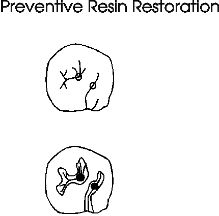
For incipient carious lesions, preventive resin restorations(PRR) should be employed. PRR is a conservative treatment that involves limited excavation to remove carious tissue, restoration of the excavated area with a composite resin, and application of sealant over the surface of the restoration and the remaining sound, contiguous pits and fissures. PRR completely eliminates the age old "extension for prevention", which is now considered as "extension for destruction".

If you are proceeding with amalgam restorations for a Class I incipient lesion, use a pear shaped bur to remove only the carious area. The cavity depth need be only 1 to 1.5 mm for a high copper amalgam restoration. Make sure you have amalgam pluggers with very small working points so that you don’t have to widen the cavity to provide convenience form. As an emergency measure, you can even cut off and flatten the tip of a straight probe to be used as amalgam condenser.
For Class II amalgam cavities, Markeley recommended a much conservative approach with a smaller box and a considerably diminished isthmus. For a very small proximal lesion, a keyway or lock across the occlusal surface is not needed. Just place retention grooves on the buccal and lingual proximal walls with a thin tapered bur.

When proximal lesions are to be restored with adhesive restorative materials, there is no need to create conventional box preparation. Instead, there are two approaches.
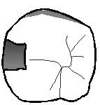
- The external "trough", "slot", or "groove" preparation. This approaches the lesion directly, either from buccal, or lingual, or from the marginal ridge.
- The second type is the "tunnel preparation", which is also known as "internal oblique preparation" "internal fossa preparation", "internal occlusal diagonal or internal preparation".

Tunnel preparation and restoration with a glass ionomer cement is possible due to the chemical adhesion of the cement, which will strengthen the weakened proximal enamel wall and marginal ridge The transference of fluoride ions offers caries protection to the adjacent tooth surfaces. Since the material allows the flow of moisture, the dehydration of the cusps is reduced, making them less vulnerable to future fracture. If there is any likelihood of occlusal load more than glass ionomer can withstand, then reduce the cement to DE junction and laminate with composite resin.
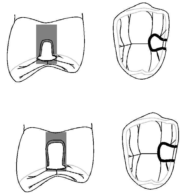
The Microchip proximal cavity preparation is a variation of the tunnel preparation when removal of the porous enamel into the marginal ridge area is required.

The mini-box proximal cavity preparation differs from the previous preparation in the handling of the enamel. Here, the integrity of the enamel wall needs to be ensured by extending the margin back to where it can be considered stable and durable.
For the gingival wall preparation, the remnants of the porous enamel is removed, and the enamel wall is taken down until stable structure is reached. This may leave the enamel wall occlusal to the dentin wall but the deficiency in the dentin can be easily restored with a glass ionomer cement.
We hope you like this info, Now click here for excellent section to learn Endodontics.If you looking for a great place to get clinical training CLICK HERE
References:
1. Louis W. Ripa and Marks S. Wolff: Quint Int. 23(5): 307-315.1992
2. Hunt PR.: JADA 120:37-40,1990.

No comments:
Post a Comment