Monday, September 29, 2008

A novel approach for treating fluorosis stains

Dental fluorosis is a common disease in punjab(india).it is due to an unusually high dose of fluorides during odontogenesis causing a structural modification of hard dental tissues and thereby resulting in a hypomineralisation of these tissues.fluorotic enamel is a hypocalcified, porous,brittle and most unaesthetic tissue. Bleaching has been suggested by several authors in order to treat the unaesthetic aspect of dental fluorosis, but many results are however unsatisfactory. This is a novel method which is based on the structural characteristics of the fluorotic ename & organic and exogenous nature of fluorotic enamel stains which includes four different stages:-

1) Cleansing the enamel surface with pumice 2) Enamel etching with hydrochloric acid 3) application of sodium hypochlorite. 4) application of dental adhesives.

Introduction

A frequent question asked by most of patients residing in the fluorotic belt of punjab (india) is "will my tooth turn white?" Usually the answer is a "yes" with the explanation that the modern dental treatment procedures are such as to esthetically synchronise the facial harmony of tooth structure.the reason for this discoloration is a high fluoride concentration in water in certain areas of punjab. the normal colour of permanent teeth is greyish yellow, greyish white or yellowish white but the number of people with this colour are usually limited owing to over aggressive tooth brushing and abrasive cleansing materials, acidic food and drinks and last but not the least,ageing. The elderly people thus, have more yellowish teeth as compared to younger persons.these alterations in colour maybe physiologic or pathologic and endogenous or exogenous in nature. the modern era is an era of esthetics. People having teeth with normal colour also want to have whiter teeth to improve their smile. So one cannot ignore the wishes of such patients and hence bleaching, as we know, has emerged as the simplest, most common, least invasive and least expensive means available to dentists to lighten discoloration.

History

Many agents have been used in the past and a number of new methods have continued to be introduced. It was oxalic acid first by chappel in 1877 which was followed by various forms of chlorine, until hydrogen peroxide was first used by harlan in 1884. Many advances continued focussing basically on the ways to facilitate the absorption of bleaching agent. The recent developments of hi-tech computer imaging have enhanced patient understanding, expectation and ultimately satisfaction.

Mode of action

Bleaching works by oxidation in which the bleaching agent enters the enamel &/or dentin of the discolored tooth and reduces the molecules containing discoloration. The bleaching depth depends on the cause of the stains and where and how deep the stain has permeated the tooth structure plus how deep the bleaching agent can permeate to the source of discoloration and remain there long enough to release deep stains.

Etiology of tooth discoloration

Extrinsic discolorations are found on outer surface of teeth and are usually Of local origin e.g. Tobacco, paan, tea, coffee, silver nitrate stains, oral intake of iron suspensions, continuous use of mouth washes and gum paint. Intrinsic the stains are found within the enamel and dentin and are caused by The deposition of the substances within these structures e.g. Tetracycline, Fluorosis stains, amelogenesis imperfecta, dentinogenesis imperfecta, pulp necrosis etc.

Histopathology

fluorosed teeth are also called mottled teeth . Such teeth appear when child ingests excessive fluoride during enamel formation or calcification in areas where drinking water contains more than 1ppm of Fluoride. The higher concentration of fluoride is believed to cause a metabolic alteration in the ameloblasts which results in defective matrix & improper calcification. 1ppm of fluoride has no biological side effects on The vital organs of human body i.e. Kidney, heart & lungs. Fluoride up to 4ppm in drinking water occasionally produces skeletal fluorosis but above 8ppm coupled with malnutrition positively causes not only skeletal fluorosis but irreversible bone changes & deformity as well.

Histology

histological examination shows hypomineralised, porous sub-surface enamel below a well mineralised surface layer. The most affected teeth (in decreasing order) are premolars, 2ndmolars, followed by maxillary incisors, canines & 1st molars. Mandibular incisors are affected least.

Stages of fluorosis

the appearance of teeth depends upon the severity of the lesion which in turn depends upon the fluoride contents consumed by a particular individual through the water supply.

1) the constant use of water having fluoride to the extent of 1ppm causes mildest grade of mottling in 10% of the population.

2) as concentration of fluoride increases, the effect worsens, so much so that when the concentration reaches 6ppm,incidence of mottling is 100%.

3) very mild :- in this type there are very small white areas occasionally seen on the tooth surfaces, but do not involve more than 25% of tooth surfaces.

(4) mild :- in this type there is more extensive tooth involvement and involves 50% of tooth surfaces.

(5) moderate :- more surfaces are involved here and are subjected to attrition. They show marked wear with yellow or brown pigmentation.

(6) severe :- all enamel surfaces are involved, so much so that the tooth mor phology is affected.there is discrete or confluent pitting of enamel surfaces. Brown stains are widespread & the tooth often presents a corroded surface.

Optimum fluoride levels

In cold climate, recommended fluoride levels may be as high as 1.2 ppm whereas in extremely hot climate, a level of 0.7 ppm is recommended. In moderate climate, the optimum fluoride level is 1 ppm. High temperature causes increase in mottling because there is increased consumption of water containing fluoride. Distribution of mottling in various areas of teeth has no relation with periods of mineralisation of crown. Teeth are only affected provided the child lives in the area of fluorosis during the time of enamel mineralisation. Brown tooth stains respond to treatment but white stains are not effectively resolved. It has been observed that teeth in process of eruption receive maximum benefit from optimum amount of fluoride plus teeth exposed to f shortly after eruption were also protected although to a lesser degree.

Different f levels in punjab & other states of india

I) Punjab

1) bhatinda - 4.5 ppm
2) mansa - 4.2 ppm
3) mukatsar - 3.3 ppm
4) faridkot - 3 ppm
5) ferozepur - 2.6 ppm
6) moga - 2 ppm
7) sangrur - 1.35 ppm
8) jalandhar - 0.55 ppm
9) amritsar - 0.45 ppm
10) hoshiarpur - 0.44 ppm
11) nawanshahar - 0.4 ppm
12) fatehgarh sahib - 0.37 ppm
13) patiala - 0.35 ppm
14) ropar - 0.3 ppm
15) kapurthala - 0.25 ppm
16) ludhiana - 0.22 ppm
17) gurdaspur - 0.15 ppm

II) Andhra Pradesh

1) nalgonda - 20.6ppm
2)prakasan - 12.0ppm
3)vishakhapatnam - 11.0ppm
4) anantpur - 10.1ppm
5)guntar - 10 ppm
6)medak - 9.8ppm
7)kunoor - 9.6ppm
8)nellore - 8 ppm
9)mehboobnagar - 6.4 ppm
10)warrangal - 5.8 ppm
11) kareemnagar - 4.9 ppm
12) hyderabad - 4.8ppm
13) cuddapah - 4.6ppm
14) nizamabad - 3.0ppm
15) chittoor - 2.9 ppm
16) adkabab - 2.8 ppm
17) srikakalam - 2.8 ppm
18) Godavari - 1.6 ppm

III) Gujarat

1) kutch - 1.2 -- 11 ppm
2) bhavnagar - 1.5 - 4ppm
3) jamnagar - 1.5 - 4ppm
4) rajkot - 2.5ppm
5) saurashtra - 1.5 - 2.5 ppm
6) rajpur - 0 - 2.5 ppm
7) banakanta - 1.5 - 2ppm
8) godar - 1.6 - 1.7ppm
9) godhra - 0 - 1.6 ppm
10) surinderanagar - 0 - 1.5 ppm
11) surat - 0 - 1.3 ppm

IV) Tamil Nadu
1) Coimbatore 2) Dharampur 3) Madurai 4) Narkot 5) Salem 6) Trichi
all 1.5 --- 5ppm

V) Kerala
1) Allepey 2) Eranakulam 3) Quillon 4) Trichur
all 0 -- 1.5 ppm

V) Rajasthan

1) Bharatpur - 28ppm
2) Tonk - 21ppm
3) Alwar - 20.6ppm
4) Sikar - 19.1ppm
5) Ajmer - 18.4ppm
6) Bhilwara - 16.5ppm
7) Swaimadhopur - 16.1ppm
8) Jhalawar - 16ppm
9) Churu - 16ppm
10) Jodhpur -16ppm
11) Sirohi - 15.8ppm
12) Jaipur - 15ppm
13) Nalpur - 14.2ppm
14) kota - 14.2ppm
15) dungarpur - 12ppm
16) bikaner - 10.2ppm
17) barmer - 10ppm
18) pali -- 9.1ppm
19) ganganagar - 9ppm
20) jalour -- 8ppm
21) wagpur - 7.1ppm
22) chittorgarh - 6ppm
23) bundi - 5.8ppm
24) banswara - 4.3ppm
25) jhunjhunu - 2.2ppm

VI) Uttar Pradesh

1) Gorakhpur - 0.6-6.8ppm
2) Shahjahanpur - 4ppm
3) Lakhpur -0.1-4ppm
4) Rai bareilly - 0.6-3ppm
5) Banda - 0.6-3ppm
6) Agra - 0.2-3ppm
7) Kanpur - 0.2-3ppm
8) Varanasi - 0.2-3ppm
9) Unna - 0.1-3ppm
10) Aligarh - 0.4-2ppm
11) Allahabad - 0.2-2ppm
12) Itah -0.8-1.6ppm
13) Hamirpur - 0.6-1.6ppm
14) Azamgarh - 0.1-1.6ppm
15) Muradpur - 1.0-1.4ppm
16) Jamalpur - 1.0-1.2ppm
17) Lucknow - 0.8-1.2ppm
18) Meerut - 0.4-1.2ppm
19) Bulandshahar - 0.4-1.2ppm
20) Dijnor - 0.2-1.2ppm
21) Jhansi - 0.2-1.2ppm
22) Bareilly 0.1-0.9ppm
23) Balliya - 0.4-0.8ppm
24) Barabanki - 0.4-0.8ppm
25) fatehgarh - 0.4-0.8ppm
26) mirzapur - 0.4-0.8ppm
27) gadhepur - 0.3-0.8ppm
28) gonda - 0.2-0.8ppm
29) basti - 0.2-0.8ppm
30) jalpum - 0.1-0.8ppm
31) dehradun - 0.1-0.8ppm
32) pratapgarh - 0.4-0.6ppm
33) manipuri - 0.4-0.6ppm
34) lahtpur - 0.1-0.6ppm
35) muzaffarnagar - 0.2-0.5ppm
36) rampur - 0.2-0.4ppm
37) pilibhit - 0.2-0.4ppm
38) bijnor - 0.1-0.4ppm
39) fatehabad - 0.1-0.4ppm
40) badari - 0.1-0.4 ppm
41) sitapur - 0.1-0.4ppm
42) saharanpur - 0.1-0.4ppm
43) mathura - 0.1-0.4ppm
44) faizabad - 0.2ppm
45) etawah - 0.1-0.2ppm
46) nainital - 0.1-0.2ppm
47) dahrich - 0.1-0.2ppm
48) sultanpur - 0.1ppm

Treatment options

Basically for all these stains or in particular fluorotic stains the treatment options available to us include :-

1) Veneering / laminates or placement of porcelain crowns
2) Micro / macroabrasion
3) Bleaching - a) vital tooth inoffice bleaching b) nightguard home bleaching c) our novel method of inoffice bleaching

1) Veneering or laminates or ceramic crowns

Advantages:
1) esthetically more acceptable
2) Long lasting
3) Durable
4) Simple
5) Can be given over endodontically treated tooth
6) more strength and resistance to forces

Disadvantages:
1) brittle
2) less shear strengh
3) causes loss of tooth structure
4) patient may not be willing
5) susceptible to fracture
6) due to tooth reduction, pulp & other tissues may face trauma
7) overcontouring may make it appear & feel unnatural
8) vitality tests cannot be done once crowns are properly fit
9) post cementation caries difficult to detect
10) lab.procedure needs precision for proper marginal seal
11) gingival irritation- may cause hyperaemia & bleeding


2) Micro/ Macro abrasion :-

This technique involves applying of 18% hcl to soften the enamel And then abrading it with a controlled abrasive technique With pumice to remove superficial stains / defects. Instead of pumice, even silicon carbide may be used with 11%hcl.

Advantages:
1) improved method for superficial stains
2) safer method
3) involves physical removal of tooth structure

Disadvantages:
1) can cause sensitivity
2) causes wearing of tooth structure
3) patients might not allow cutting of tooth structure
4) defect may persist after finishing of technique for which a restorative alternative is needed


3) Bleaching :-

This procedure has many methods and techniques involving various solutions in each technique.

Advantages:
1) easy
2) time saving
3) cheaper
4) patient acceptance better
5) can be carried out both in office & at home

Disadvantages:
1) requires patient cooperation(especially for home bleaching)
2) cannot be used where teeth have large pulps
3) cannot be used where teeth are too dark
4) cannot be used where the patient expectations are too high
5) cannot be used in impatient patients
6) causes cervical resorption
7) cannot be used in attritioned teeth which might cause sensitivity
8) cannot be used where teeth are bonded, laminated or have extensive restorations
9) not a perfect technique & merely changes colour to variable depths
10) lasts for only 1 - 3 years (short period)

A) Vital tooth inoffice power bleaching
This technique uses a combination of 37% phosphoric acid & 35%hydrogen peroxide.the oxidation reaction is generally promoted by a heated instument or with intensive light.in this method, one application is carried out weekly for 2 - 6 appointments with each treatment lasting 30 minutes. Use of phosphoric acid by this technique is optional.

Advantages:
1) caustic chemicals are totally under dentist's control.
2) soft tissue protection is better achieved by dentist.
3) bleaching of tooth is achieved more rapidly

Disadvantages:
1) slightly costly procedure.
2) unpredictable results.
3) uncertain duration of treatment
4) soft tissue damage possible for both dentist & patient.
5) rubber dam causes discomfort.
6) can cause post operative sensitivity.

B) Night guard home bleaching
This procedure involves making an impression of the teeth & pouring a cast of the same, trimming of the cast, application of a blockout resin & fabrication of a night guard tray by a vaccum former machine. After cooling, the tray is trimmed & a 10 - 15% gel of carbamide peroxide is recommended for the same. In this procedure the total treatment time is 2 - 6 weeks.

Advantages:
1) use of lower concentration.
2) ease of application.
3) minimal side effects.
4) lower cost (as compared to veneers)
5) lesser chair time.
6) much lesser labour intensive.

Disadvantages:
1) have to rely a lot on patient compliance for results.
2) longer treatment time.
3) unknown potential for soft tissue changes with excessive use.
4) treatment results are time & dose dependent.
5) peroxide solution may cause irritation of gingival papilla.
6) teeth become sensitive to temperature changes.

Another method using macken's solution has been described
1 part anaesthetic ether 0.2 ml - removes surface debris 5 parts hcl 38% 1ml --- etches 5 parts hydrogen peroxide 30% 1 ml --- bleaches


Our Approach For Inoffice Bleaching

Indications:
1) Fluorosis stains / systemic fluorosis
2) Tetracycline stains

Contra indications:
1) Hyperaemic gingiva
2) Persistant periodontal problem cases
3) Fractured incisors / anteriors

Clinical application
The various steps are
1) Cleansing
2) Isolating
3) Etching
4) Rinsing
5) Dehydration
6) Application of solution
7) Scrapping
8) Rinsing
9) Filling

The Steps in detail:
1) cleansing the tooth surface with a nylon tooth brush & a mixture of pumice and water to remove surface debris.
2) isolation is done by application of rubber dam.
3) then dry the tooth surface & do enamel etching with 35% hcl for 20 - 25 seconds.
4) copious rinsing is done to eliminate acid residues & the tooth is subjected to thorogh drying.
5) application of 95% ethyl alcohol to dehydrate the enamel surface.
6) now,the application of 30% hydrogen peroxide(h2o2) is done first for 1 minute followed by alternative application of 5.25% sodium hypochlorite (naohcl) is done for 5 minutes during which it can be re-applied to the tooth surface to keep it wet.
7) the removal of staining molecules can be accelerated by gently scrapping the tooth surface.
8) this is followed by thorough rinsing of tooth surface.
9) this procedure is repeated at the interval of three days for successive sittings till the results are satisfactory.
10) in the end, fill the microcavities caused in the tooth by this solution with a light cure dental adhesive.

Advantages:
1) HCl etches enamel,but does not penetrate.
2) Tooth structure is not damaged.
3) Very very few chances of post - operative sensitivity of tooth.
4) No heat / application is required.
5) Very economical as all the three solutions in quantity of 50 ml. Each cost rs. 250 - 300 (total ).
6) Very low quantity of solutions required at each sitting.

Disadvantages :
1) Fluorosed teeth require larger & repeated sessions to decolorise Them.
2) Some blanching of gingiva can occur which is reversible within Half an hour.
3) Transitory decrease in bond strengh occurs when composite is applied to bleached / etched enamel.however,after a week,no decrease is seen.
4) Unknown duration of treatment.

Discussion

The different hypothesis concerning the fluorotic stains removal are:
1) if a fluorotic tooth is put into a naohcl solution,it removes all the stains within a few hours.this confirms the organic & exogenous nature of fluorotic tooth stains which are due to elementary impregnation of a hypocalcified & porous tissue. said by :- triller m. Alterations des tissues by marie curie in 1984.
2) scanning electron microscope study (sem) study shows that Posteruptive calcified layer covers the fluorotic enamel surface ; hence the mineral layer removal is essential.

Conclusion

In the end, i would like to conclude that this system of stains Removal seems to be clinically applicable & satisfactory with minimal abrasion of enamel surface.to make this technique Universally acceptable , lot of cases have to be treated with this technique.

Immediate and Early loading of Implants

Protocols for immediate or early loading strive for an increase either of primary stability, which can be achieved either through an optimized implant form or implant surface or/and optimized surgical preparation of the implant bed. Conversely, through the modification of the implant surface, an acceleration of the bony healing is intended to achieve an earlier osseointegration and therefore a faster acceptable secondary stability for successful loading. The authors conclude that the use of immediate loading still consists of a higher risk because of non-existing, not yet defined diagnostic criteria and can therefore not be recommended to the general practitioner. An acceptable measurement system may be available in the near future and help revolutionize the treatment concept in implant therapy.


Osseointegration is the result of a biologic response of the bony tissues to implants. For oral endosteal implants a three to six month healing period prior to loading is generally considered a pre-condition for optimal bone apposition to the fixture. This clinical protocol is based on work done by Branemark during the 1960's and 1970's when a final proposition for healing periods for implants of the upper and lower jaw, based on the evaluation of the surgical and periodontal failure rates, was defined (Brånemark et al, 1977 [1] ). These protocols on Osseointegration periods were based on early, partially unfavorable experimental conditions while methods and implant types were continuously changing:
  • On edentulous patients with a reduced bone quality and quantity

  • With short implant lengths

  • With changing less than optimal implant designs

  • With changing less than optimal surgical protocols

  • With not optimized biomechanically suprastructures


It is therefore not surprising that in the past, the submerged healing period was frequently questioned, based on modifications of surgical and prosthetic procedures. In several animal and human studies the original three to six month healing period was shortened to an early loading protocol. In other modifications, implants were immediately loaded on the day of insertion.


Post-Surgical Bone Reaction
Concerning the bone healing after the surgical implant placement, one can draw the following conclusion:

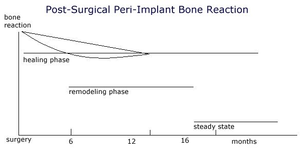
The perimplant bone reaction is initially the highest after the surgical trauma and reduces itself slowly over months, returning to the basic turnover. It is evident that even after the three to six month standard healing protocol any loading will not be placed on the final bony implant bed but bony remodeling will still be in progress.


Based on these two conditions it is not feasible to determine a general healing period depending only on the type of jaw. To define an individual healing period the bone should be evaluated both during and after the implant surgery. Because traditional methods are unsuitable for this measurement, new methods are recently described.

Methods for the evaluation of the peri-implant bone conditions:
Traditional methods
  • Percussion tests (Adell et al. 1985 [2])

  • X-ray evaluation (Lekholm & Zarb 1985 [3])

  • Periotest (Olivé & Aparicio 1990 [4])

  • Turn out tests (Sullivan et al 1996 [5])

Modern methods
  • Cutting resistance measurement during implant placement (Johansson & Strid 1994 [6])

  • Resonance frequency analysis (Meredith et al. 1996 [7])


The recording of the cutting resistance during the surgery permits some evaluation of the bony structure (compact versus spongious) along the implant site. The measurement of the resonance frequency analysis on an inserted implant provides information about the primary stability between implant and bone. While primary stability in implantology was previously based on a purely subjective evaluation, these two new measurement methods permit an objective recording of the initial implant stability.


Primary and secondary implant stability
To achieve osseointegration an attachment to the bone without any micro-movement is necessary. Prior dental literature cites micromovement of the implant as a causative factor in both the formation of an intermediate layer of connective tissue, which develops between the bone and implant, and osseointegration failure. Initially, primary stability is entirely mechanical. During the healing period however, the biologic processes of osseointegration cause this to change to a mixture of mechanical and biologic stability (secondary stability).

Protocols for immediate or early loading strive for an increase either of primary stability, which can be achieved either through an optimized implant form or implant surface or/and optimized surgical preparation of the implant bed. Conversely, through the modification of the implant surface an acceleration of the bony healing is intended to achieve an earlier osseointegration and therefore a faster acceptable secondary stability (Case examples Fig. 1 - 12).

Figure 1
Figure 2
Figure 3
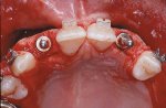
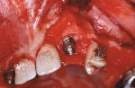
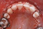
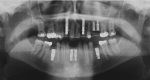
A 19-year-old patient, with multiple genetically missing teeth, at the end of the preprosthetic orthodontic treatment. In the upper jaw implants are planned in the area 12 - 25. Implants on 12 and 22 should be immediate loaded because of esthetic reasons.

Figure 4
Figure 5
Figure 6
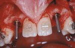
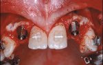
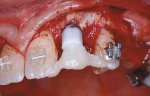
After preparation of the implant bed at the position of both lateral incisors the prospective implant placement will be marked with a direction indicator. Two Brånemark MK IV are inserted and a CeraOne™ abutment is definitively placed on both implants.

Figure 7
Figure 8
Figure 9
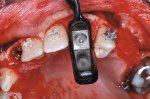
Initial implant stability will be measured with the RFA method. The buccal bony defect will be covered using a membrane technique and a prefabricated shell-like temporary will be directly, intraorally rebased over the abutment.

Figure 10
Figure 11a
Figure 11b

Figure 12


Summary
The protocol of successful osseointegration, which was based for over 20 years on the concept of late loading, is increasingly questioned. In several studies successful immediate loading or early loading of implants could be shown. Two indications were described:
  • Bar-joint-dentures (Dolder bar) in the lower jaw on 4 - 6 interforaminally placed implants

  • Total bridge in the upper or lower jaw on 10 or more implants


The indication for the case selection for immediate loading is based on an optimal stabilization of the implants through a large number (full, horse shoe type bridges) or on implants in a very good bone quality or quantity (interforaminal). Through secondary splinting with the suprastructure, possible micromovement during the healing period can be prevented in both situations.

All protocols up to the present day however show that the operator based case selection on subjective criteria. Objective quantitative criteria for case selection are not yet published. Long term data on immediate, or early implant loading exist only for interforaminally placed fixtures.

At the moment the use of immediate loading still consists of a higher risk because of non-existing, not yet defined diagnostic criteria and can therefore not be recommended to the general practitioner. On the other hand a suitable measurement system will be available in the near future and might revolutionize our treatment concept in implant therapy.

Options for esthetic restorations

Contemporary dental practices have embraced the process of bonding to natural tooth structure to combine function and esthetics. Direct composite resin restorations have replaced amalgam and gold in many clinical situations where esthetics is of primary concern and an adequate amount of sound natural tooth structure remains.

When more extensive reconstruction is required or elective treatment requires covering larger areas of the tooth, indirect laminates of processed composite resin or ceramic materials can be used. A laminate can cover only the facial surface of a tooth or it can wrap around to inter-proximal or lingual areas where esthetics or function demands more aggressive treatment. Where conventional treatment may call for a full crown, a 360-degree laminate can accomplish the same result while requiring a thickness of as little as 0.5 mm.

While ceramic materials are inherently brittle and can be broken easily when unsupported, they become quite strong when laminated to an underlying durable material. The lamination process is why porcelain fused to metal has been so successful in dentistry. The fact that the porcelain is laminated to a metal understructure gives it the strength to hold up under heavy functional stresses in the oral cavity.

Now that we can bond ceramics and composite resins to natural tooth structure, the tooth itself becomes the supporting structure and no metal foundation is needed for many clinical situations. Because there is no need for metal and an opaque layer of porcelain to block out the color of the metal, the restoration can be thinner so less tooth reduction is required. More of the patient's natural tooth structure can be maintained.

Restorative Materials

But as we communicate these facts about advances in materials to our patients, we must temper our enthusiasm and provide balance. The word "permanent" should be eliminated from our dental vocabulary. Even natural teeth wear away as we age. Gold is the closest thing to permanent we can use for dental reconstruction. However, today most patients prefer tooth-colored alternatives.

Bonded composite resin and ceramic materials can deliver the desired result while providing longevity that is acceptable to most patients. Still, it is very likely that the restoration a patient receives today will need replacing sometime in the future. While most dentists agree that bonded ceramics provide greater longevity than composite resin, ceramic materials are more difficult to remove.

Composite resin can be removed with carbide finishing burs and it is easy to detect when natural tooth structure is reached, so little or no additional tooth removal may result. Porcelain, however, must be ground off using diamond burs and it is very likely that more tooth structure will be removed at each new preparation. I prefer to prepare conservatively so the patient will have as much natural tooth structure as possible left for future treatment.

Direct composite resin restorations require the least tooth reduction since they are built directly on the prepared tooth. They can be paper-thin in areas and still perform well. Laboratory-fabricated processed composite resin restorations need slightly more preparation because they must have a certain thickness (as little as 0.3 mm) to be held in the technician's hand and finished without breaking. Stacked porcelain needs only a little more thickness (0.3 to 0.5 mm) but still can be very thin and function well after being bonded to the natural tooth. Pressed ceramics require more aggressive tooth reduction (0.6 mm and up) because they are fabricated using the lost wax technique. The process simply requires more thickness to allow for fabrication of the ceramic core and overlaying of low-fusing porcelain for esthetics.

The Lamination Process

Regardless of the material chosen, the bonding process requires attention to detail and flawless clinical technique. There is no span between excellent and poor in adhesive dentistry. If the clinical result is not excellent, it is no good at all. Inadequate bonding can lead to marginal leakage, tooth sensitivity, recurrent decay, de-bonding and loss of the restoration.

Current science advocates wet bonding. Our modern bonding agents are hydrophilic, allowing for bond strengths on dentin and enamel that exceed the cohesive strength of both the natural tooth and the restorative material. The luting composites for indirect laminates of composite resin and ceramic materials are of major importance in the success of the restorations. Indirect inlays, onlays and full crowns rely on composite luting agents for their retention and marginal seal.

The bonding process actually results in a strengthening of the remaining natural tooth structure since the remaining parts are held together instead of being wedged apart, as was the case with amalgam fillings.

What About Sensitivity?

Post-operative sensitivity has been one of the major areas of concern for most dentists who perform adhesive procedures for their patients. While some clinicians have claimed no problems with sensitivity, the vast majority of dentists are concerned about this phenomenon. Cold sensitivity is the most common complaint. This can occur due to open margins and resultant micro-leakage. However, even dentists who are sure that a marginal seal has been achieved have expressed frustration that cold sensitivity can still exist.

The most likely explanation is that a hybrid layer has not been achieved in the dentin bonding process or there are open dentin tubules which have not been sealed. By acid etching the enamel for 15 seconds with 37% phosphoric acid, part of the enamel matrix is dissolved, leaving thousands of tiny micro-pores in the enamel surface.

A liquid resin is flowed into these pores and allowed to harden, forming thousands of fingers of resin into the enamel. The bond is tremendous and is stronger than the tooth. The same acid removes the smear layer from the dentin in just five seconds, leaving the dentin tubules open. A hydrophilic resin flows into these open tubules and into the inter-tubular dentin, forming a "hybrid" layer consisting of resin and dentin which is mechanically and chemically locked together at a strength which is higher than the tooth itself. If this hybrid layer is incompletely formed or absent, the dentin tubules remain open and a negative pressure results when temperature or osmotic changes occur in the oral cavity. This results in the sensation of pain.

Given enough time, cold sensitivity usually goes away. I believe many endodontic procedures have been prematurely performed by well-meaning dentists who did not understand this process. However, with proper technique, post-operative sensitivity can be nearly eliminated today.

A dentin desensitizer and wetting agent used after acid etching and before applying the bonding agent can be very beneficial in helping form this important hybrid layer. Several formulas are currently on the market. One of the popular brands contains gluteraldehyde as its anti-microbial agent. This chemical can tan gingival tissues, so it must be used carefully in order to confine it to the tooth structures. I prefer another well-known brand (Hurriseal, Beutlich Pharmaceuticals) that uses benzalchonium chloride as the anti-microbial agent. It is kind to all tissues and can be used anywhere in the oral cavity.

Bonding Agents

Fourth-generation bonding agents utilized a primer followed by an adhesive. Fifth-generation bonding agents combine these into one solution. Both can work effectively when used properly. Film thickness is the major concern for one-step bonding agents since they must be light cured before a luting agent is placed between them and the restoration.

Many current one-step bonding agents exhibit a very low film thickness and can be used successfully for indirect procedures. When in doubt, a fourth- generation bonding agent can be used since it is cured only after the restoration is placed.

Luting Agents

Luting composites can be either microfills or micro-hybrids. Handling properties usually determine the choice by the clinician. Some are less viscous and flow readily. Others exhibit a higher viscosity. I like the higher viscosity resins and find clean-up procedures less difficult with their use.

Luting composites can be light cured or dual cured. I find dual-curing resins more efficient to use as long as a distinct gel state occurs during the curing process. At this stage (approximately three minutes after the start of the mix of the catalyst and base for most materials), the excess can be easily peeled away with a gentle touch of a scaler or explorer. Nearly total clean up can be accomplished before light curing.

For those who are concerned about stresses that may be caused by high-speed curing, the dual-cure technique will eliminate this problem. Some clinicians have expressed concern that dual-cure resins can darken with time, as did self-cure resins of the past. I must say that I have never seen this phenomenon clinically and I have been using dual-cure luting agents routinely for years.

Finishing procedures

It is hypothesized by some that sensitivity is created by aggressive finishing procedures. Care should be taken to minimize use of rotary instruments at the marginal areas where dentin and cementum are present. Careful technique must be used with light-cured luting resins. You must be sure to remove as much excess resin as possible before light curing. This will minimize the need for finishing. By using a dual-cure resin, most of the excess can be removed before light-curing, so post-cure finishing is automatically minimized. Of course, excellent margins that are clinically undetectable and flow into the natural tooth anatomy allow for maximum dental health and reduce the likelihood of any post-operative problems. Esthetic supra-gingival margins are usually possible and preferable when metal-free restorations are used.

Conclusion

The choice of direct or indirect composite resin, stacked porcelain or pressed ceramic materials depends on many factors. When considering the restorative material, conservation of tooth structure will often lead to the correct choice. Sometimes other considerations, such as the presence of old restorations or broken down tooth structure, may dictate or allow the use of materials which require more aggressive preparation.

A desensitizer and wetting agent can help eliminate post- operative sensitivity and ensure a complete hybrid layer. The use of a fourth- or fifth-generation dentin and enamel and bonding agent will assure bond strengths that exceed the cohesive strength of the tooth structure or the restoration. Modern luting composites can be light cured or dual cured. The dual-cure technique can result in easier clean up and less need for marginal finishing which may also result in less post-operative sensitivity.

A primer on all composite class materials

Direct and Indirect Esthetic Adhesive Restorative Materials : A Review


Dr। Kugel is Professor and Assistant Dean for Research, School of Dental Medicine, Tufts University, Boston, Massachusetts, USA


** Dr. García-Godoy is Professor and Director, Clinical Materials Research, Department of Restorative Dentistry, University of Texas Health Science Center at San Antonio, Texas, USA.

Esthetic direct alternatives to amalgam restorations include glass-ionomers, resin-modified glass-ionomers, compomers and composite restorations. Marked changes in the use of restorative materials have occurred during the past 10 to 20 years1-5 and esthetic considerations are growing in importance for the restoration of posterior teeth.6 Alleged adverse health effects and environmental concerns due to the release of mercury launched controversial discussions about the continued use of amalgam as a contemporary restorative material.7,8

In recent years, development and interest in esthetic dentistry has dramatically increased. This is evident from dental journals publications. Many of the articles and an increasing number of entire journals are devoted to this topic. Along with the journals comes an avalanche of advertisements with products claiming to be the new breakthrough in esthetic restorative dentistry although many times there is nt enough clinical evidence to support their use. The patient population is not immune from this onslaught. They read about bleaching, lasers and metal-free dentistry while standing in line at the supermarket. These same patients are living in a youth oriented society, which places a high value on appearance. Add to this the unsupported fear of mercury in dental amalgams and is it any wonder that the dental professionals are flocking to educational institutes and seminars all across America in search of the esthetic revolution.

As amalgam replacements have become a primary focus of dental materials research, the need for improved adhesion has also become an area of major interest. The principles of adhesive dentistry date back to 1955 when Buonocore,9 employing techniques of industrial bonding, suggested that acids could be used as a surface treatment before applying resins. In the late sixties Buonocore et al10 believed that resin tag formation was the cause of adhesion to acid etched enamel. This principle is well accepted today.

As we advanced in our understanding of enamel and dentin bonding we also sought to improve those materials we used with our new bonding systems. We marched from our first generation of adhesives, which yielded bond strengths of only 1-3 MPa with poor clinical results, to the present use of fifth and sixth generation systems.11 For example, ESPE's recent entry into the bonding arena is Prompt L-Pop. This is a sixth generation self-etching adhesive system that incorporates the etching of enamel and dentin in one application without a bottle (“bottleless” adhesive).12

Figure 1

(A) Pre-op photo, teeth 12&13,
(B) Preparation,
(C) Prompt-L Pop Bonding System,
(D) Immediate post-op, ESPE’s experimental fluoride-releasing composite,
(E) 6-month post-op,
(F) I-year post-op.

Esthetic restorative materials require a bonding procedure in order to be durable and reliable. In order to accomplish this, the bonding system must be biocompatible, bond similarly to both enamel and dentin, have sufficient strength to resist masticatory forces, have mechanical properties close to those of tooth structures, be resistant to degradation in the oral environment and easy to use.

The longevity of dental restorations is dependent upon many different factors including materials-, dentist-, and patient-related factors. Main reasons for failure are predominated by the formation of secondary caries, fracture of the bulk of the restoration or of the tooth, marginal deficiencies, and wear.

The importance of direct placement esthetic tooth-colored restorative materials is still increasing. Direct composite restorations require a time-consuming and more costly treatment procedure.

Our laboratories reported the benefit of using resin-modified glass-ionomers (RMGIs) as liners under posterior composites and amalgams as a way to reduce cervical microleakage13,14 and recurrent caries.15,16 This may be due to their fluoride release.17,18 Glass-ionomers have also been shown to be antibacterial.19

From an esthetic standpoint, glass-ionomers can only be considered as "long-term" provisional restorations in stress-bearing posterior cavities and will not be addressed in this review

Direct Adhesive Restorative Materials

The availability of fluoride-releasing dental materials have been increasing and the following outline highlights the amount of fluoride release:

Fluoride Releasing Restorative Fluoride Release

Conventional glass-ionomer High

RMGI -->High

Smart composite --> High-Med

Compomer --> Medium

Ceromers --> Low

Composite Resin --> Low

Packable composite --> Low

Flowable composite --> Low

Ormocer --> Low

Composite Resin

Along with improved bonding the clinical performance of direct and indirect restoratives has improved. The most commonly used direct esthetic restorative material is composite resin. The clinical performance of these materials has improved due to developments in wear resistance, color stability, strength, radiopacity and percent cure.20 There are three main factors affecting the properties of composite resins: they are the filler, resin, and interface characteristics. The fillers in composite resins are added to control their handling characteristics, reduce shrinkage, increase strength and decrease abrasive wear.

The fillers used include:

  1. Amorphous silica (microfiller)

  2. Quartz

  3. Radiopaque glasses, barium, strontium, others

  4. Sol-gel zirconia-silica

  5. Fluoride containing fluorosilicates, ytterbium and yttrium trifluoride

(A) Pre-op photo, teeth 12&13, (B) Preparation, (C) Prompt-L Pop Bonding System, (D) Immediate post-op, ESPE’s experimental fluoride-releasing composite, (E) 6-month post-op, (F) I-year post-op.

One of the major improvements in composite resin has resulted from increased filler loading along with varying its distribution, size, shape and composition.

Table I - Filler Classification
TypeAverage (mm)Large (mm)Vol %
Microfill0.04 - 10.1 - 5.038 - 50
Minifill0.4 - 0.81.0 - 5.056 - 66
Midifill1.0 - 3.05.0 - 15.070 - 77
Conventional5.0 - 15.050 - 7060 - 70

The microfilled composite resin direct restoratives are highly polishable but are subject to increased marginal breakdown.21 Most of the mini-filled composites have a small amount of amorphous silica added to the monomer to improve the handling characteristics. Midifilled composite resins employ zirconium silicate, strontium, or barium fillers and they have an average particle size of 1-3 µm (Table 1). Along with particle size, one must be concerned with filler content. Filler content is the quantity of filler in a composite. It has been demonstrated that filler volume and filler load correlate with the strength of the composite resin. Increased filler volume results in an increase in fracture, toughness (Figure 2), along with an increased flexure modulus22 (Figure 2). The wear of composite restorations depends on filler particle size, interparticle spacing, and filler loading.23 There is reduced wear with smaller particles due to less plucking and therefore fewer voids. Smaller interparticle spacing allows for matrix protection by having the matrix areas smaller than the abrasive particles.

Figure 2

The filler particles of composite resins are silanated in order for the hydrophilic filler to bond to the hydrophobic resin matrix. The importance of silanation is significant. Good silanation will result in a stable composite resin that is resistant to wear with a homogenous composition.24

The matrix of most composite resins is a mixture of di-functional monomers containing Bis-GMA, TEGDMA, or UDMA in some combination. More recent materials such as the ormorcers (organically modified ceramic) employ multi-functional methacrylates.

Flowable Composite Resin

Also included in the category of composite resins are the flowable composite resins. These resins have lower filler volumes than the conventional direct composite resin restorative materials. As a result of this lower filler volume, these materials have a decreased viscosity, which makes them a good choice as a pit and fissure restorative. However they also have increased shrinkage and increased wear along with decreased strength. They have been recommended as a class V restorative or as a liner under posterior composites.

In vitro evidence does not support their use as a liner in order to reduce microleakage.25

Recent Composite Resin Technologies

Figure 3

Compomers

Compomers are defined as polyacid-modified resins. These materials are basically light-cured, low fluoride releasing composite resins. The difference between compomers and composites is that the compomers monomers contain acidic functional groups that can participate in an acid/base glass ionomer reaction following polymerization of the resin molecule. The term compomers is the result of the hybridization of the words composite and glass ionomer. However, they are not glass-ionomer materials. A true glass-ionomer must be a two-component system or else the acid/base reaction would take place immediately.24 Conventional glass-ionomers must be mixed just prior to use. With the compomers, there is a single component system which cannot contain any water in order to prevent a premature glass-ionomer reaction.

A resin polymerization takes place with the compomers after the material has set completely. The glass-ionomer reaction may then occur in the presence of water. Water is a necessary medium for the acid/base reaction. In the presence of water from the oral cavity, the acid functional groups, which are attached to the monomer units, and have now become part of the polymerized material are able to react with the base (glass) to stimulate the glass ionomer reaction. Fluoride is released as a result of this reaction. The level of fluoride release from the compomers is significantly lower than what is seen for conventional glass-ionomers or RMGIs.26 When they were introduced, acid etching was not required by the manufacturers. This was seen as an advantage over the use of composites along with the suggested fluoride release. It was subsequently demonstrated that the use of an acid etched procedure significantly improves both the retention and marginal leakage of the compomers.27-29

In summary, compomers:

  • Consist of a paste containing Ca, Al, F silicate glass filler in dimethacrylate monomers with acrylic acid like molecules;

  • Are set by polymerization of C=C of methacrylate (delayed acid/base reaction between glass and acid molecules);

  • Promote adhesion to teeth, mediated by the adhesive.

  • Offer good strength, biocompatible, low solubility

  • Have higher wear than composite, lower F release than conventional glass-ionomers and packable composites.

Packable Composites

As the dental profession searched for an amalgam substitute it found limitations in the composite materials on the market. These limitations included: resistance to wear, fracture of the restoration within the body and at the margins, marginal leakage due to polymerization shrinkage and the technique sensitivity including the ability to obtain adequate proximal contact in the final restoration.

Packable or "condensable" composites have recently been introduced to the profession as an amalgam substitute. They contain a higher filler load as well as filler distribution. This results in a stiffer consistency than hybrid composites. These changes in the properties of the packable composites result in improved handling characteristics, at least as seen by the manufacturers of these products. The major advantage to these materials is that they make it easier to establish inter-proximal contacts while placing class II restorations.

Some of these materials also claim that they may be cured in bulk. Recent studies have indicated that some of the packable composites claiming bulk fill to 5 mm depth fall short of these claims.

Figure 4

One study using a halogen light demonstrated at a 40-second cure that the uncured values are 39.08% for Prodigy, and 39.61% for Surefil, both of which claim to be bulk fill materials. The results for the two materials, which recommend incremental cure, were 33.76% for P60 and 33.19% for Tetric Ceram30 (Figure 4).

Bingham JDR 1518A:2000

Ceromers

The term ceromer stands for Ceramic Optimized Polymer and was introduced by Ivoclar to describe their composite Tetric Ceram. This material consists of a paste containing barium glass (< c="C">

Ormocers

Ormorcer is the acronym for Organically Modified Ceramics. This class of material represents a novel inorganic-organic copolymer in the formulation that allows for modification of its mechanical parameters. The inorganic-organic copolymers are synthesized from multi-functional urethane- and thioether(meth)acrylate alkoxysilanes as sol-gel precursors. Alkoxysilyl groups of the silane permit the formation of an inorganic Si-O-Si network by hydrolysis and poly-condensation reactions. The methacrylate groups are available for photochemical polymerization.3,31 The filler particles are 1-1.5 µm in size and the material is 77% filler weight and 61% filler volume.

"Smart" Composites

This class of composites was introduced as the product Ariston pHc in 1998. Ariston pHc is an ion-releasing composite material. It releases fluoride, hydroxyl, and calcium ions as the pH drops in the area immediately adjacent to the restorative material. The drop in pH values is the result of active plaque which results in a corresponding increase in the release of functional ions.3 Smart composites work based on the newly developed alkaline glass filler which will reduce secondary caries formation at the margin of a restoration by inhibiting bacterial growth. This results in a reduced demineralization and a buffering of the acid produced by caries forming microorganisms.31

The paste consists of Ba, Al, and F silicate glass filler (1 µm) with ytterbium trifluoride, silicon dioxide and alkaline Ca silicate glass (1.6 µm) in dimethacrylate monomers: it is filled 80% by weight and 60% by volume. The use of an adhesive to tooth is not recommended. However dentin should be sealed to reduce sensitivity. The fluoride release from this material is claimed by the manufacturer to be lower than conventional glass-ionomers but more than that of compomers.

Indirect Adhesive Restoratives

In an effort to address the disadvantages of the direct adhesive restoratives such as technique sensitivity, anatomic form, polymerization shrinkage, wear and interproximal contacts, the indirect composite resins were introduced. These materials were also seen as having advantages over PFM and all ceramic restorations. PFM restorations require a metal substructure, although reliable and time-tested, they are often considered as esthetic as the all-ceramic or indirect composite restorations. It is difficult to obtain the translucency seen in natural teeth when using a metal substructure.

While the all-ceramic restorations are more translucent than PFM, they have other problems, such as abrasion of the natural dentition and they are not easily repaired.

First Generation

The first generation of the composite–based indirect adhesive restoratives were introduced in the early 1980’s. These included Visio-Gem (ESPE) and Dentacolor (Kulzer) followed in 1987 by Concept (Ivoclar). These materials were developed in an effort to overcome the polymerization shrinkage and accelerated wear experienced with the direct adhesive restorative materials. These new materials offered the advantages of chairside repair, esthetics, improved anatomy and interproximal contact along with ease of fabrication. However, this first generation of materials suffered from low flexural strength, low modulus of elasticity and low resistance to wear abrasion. These poor physical properties were the result of low filler load and high matrix load.

Second Generation

It was not until the mid-1990’s that the second generation of indirect composite restorative were introduced. These new materials included Artglass (Heraeus-Kulzer), BelleGlass HP (Kerr), Targis (Ivoclar), Colombus (Cendres et Mataux) and more recently, Sinfony (ESPE).

Figure 5

The effect of heat curing these newer composite materials on the degrees of conversion was demonstrated by Ferracane and Condon in 199232 (Figure 5). They demonstrated that heat treatment, in addition to a 60-second light cure, will increase the degree of conversion. This increase in degree of conversion will improve the fracture toughness of the indirect composite materials (Figure 6).

Figure 6

This second generation of materials incorporated ceramic fillers with mean particle sizes of less than 1 µm of smaller diameter, silanized and with a narrow distribution. The filler is commonly barium silica. They have a high filler load (70-80% by weight and 50-60% by volume) and they have a lower resin content (approximately 33% resin matrix by volume.) As discussed earlier, the filler volume and filler load correlate with the strength of a composite resin.22 The higher filler load will help reduce the polymerization shrinkage, while it increases the modulus of elasticity.

Figure 7

Wear data from the manufacturer is reported as being quite low. BelleGlass is reported by the manufacturer as having wear of 1.2 µm for 1 year. The CRA in 1998 reported their findings as being quite different. Belleglass was shown to wear at 62 µm per year, Targis, 106 µm, and Artglass, 77 µm (Figure 7).

In an attempt to expand the use of these materials, Kulzer attempted to bond their polymer glass, Artglass to a metal substructure. The results were less than satisfactory. Depew and Sorensen33 demonstrated that of 49 units placed, at year 1, 32% failed and at year 2, they had a 39% failure rate. Ninety percent of these failures occurred by the composite veneer debonding from the metal. They also reported mean maximum wear depth of 113 µm (SD56-480 µm). Their conclusion was that Artglass to metal is unreliable with high failure rates and wear. More recently ESPE has entered this arena with their product, Sinfony. This composed of 48% monomers, 1% initiator, 40% ultrafine glass, 5% pyr. silica acid, 5% glass-ionomer and 1% silane. This material shows promise but needs further evaluation.

Fiber-Reinforced Substructure

This second generation of indirect composite resins may be combined with fiber-reinforced substructure. This technology was used for periodontal splinting as well as temporary bridge reinforcement to its introduction as a reinforcement for indirect composite restoratives.34 The fibers used for this purpose are composed of carbon Kevlar, polyethylene and glass fibers (Table 2).

Fibre-Reinforcement Materials
ProductMaterial
Connect - KerrWeave, polyethylene (PE)
DVA - Dental VenturesTufts, PE
Fibreflex - BiocompTufts, Kevlar
FibreKor - Jeneric/ PentronUnidirectional, Glass
FibreSplint - Inter DentalWeave, Glass
GlasSpan - GlasSpanWeave, Glass
Ribbond - RibbondLock-stitch, Woven PE
Splint-It - Jeneric/ PentronWeave, Glass
Vectris - IvoclarWeave, Glass
Table 2

These fibers may be preimpregnated with resin. The preimpregnated fibers are silanized and coated with resin. This will provide a cohesive bond to the resin matrix.

Indications for indirect composite restoratives are:

- Metal free dentistry

- Esthetics

- Decreased wear of opposing dentition (as compared to porcelain)

- Conservative tooth preparation35

Contraindications include:

-Bruxism/clench

-Opposing porcelain

-Long span fixed partial dentures

-High caries rate

-Difficult moisture control during adhesion

The CRA Newsletter reported in 1998 surface degradation of the initial polish with significant wear rates. They also noted high levels of post insertion sensitivity. Finally, delamination from the fiber reinforcing substructure due to layering techniques used during the fabrication process have been reported.36,37

Ceramic Adhesive Restorative

This group of materials include the press ceramics such as IPS-Empress, OPC, Finesse, Creepers and VitaPress along with the reinforced core materials such as In-Ceram and Procera. These offer improved esthetics and strength but they are more difficult to fabricate and they cannot be repaired. The additional issue arises that the reinforced core materials cannot be bonded. In addition to the pressed and reinforced ceramics, there are the ceramic adhesive restoratives that are ground from preformed blocks. These are machined by use of computer-aided design/computer-aided manufacturing (CAD/CAM) or by a copy milling unit.

Conclusions

There is a plethora of direct and indirect adhesive restorative materials on the market. The average dentist is often overwhelmed by the choices. Many times one material offers little advantage over another. However, as the push for improved esthetics continues to grow, so will our list of materials. So long as there is a market, the manufacturer will introduce new products.

As dentists we must not only rely on the materials but on our own ability to treatment plan and diagnose based on a multifactorial situation. We must be careful in our preparation, design, communication with a skilled laboratory technician and be meticulous in our bonding techniques. Failures in dentistry are frequently blamed on the materials. In reality, “user error” is often the culprit. Interpreting the research data and understanding our options based on the patients’ caries risk status are the first steps toward success.

Fiber Reinforced Composites

The technology and materials available to today's restorative dentists offer various solutions to many complex problems. Missing tooth structure can be replaced through the use of adhesives or metal-ceramic crowns, and missing teeth can be replaced with any of a variety of fixed prostheses supported by teeth or implants. Porcelain-fused-to-metal substructures continue to be a mainstay of fixed prosthodontics,and polymethyl methacrylate (PMMA) polymer remains the material of choice for complete denture bases. But we now also have a new emerging material that is Fiber Reinforced composites (FRC).

Fiber-reinforced materials have highly favorable mechanical properties, and their strength-to-weight ratios are superior to those of most alloys. When compared to metals they offer many other advantages as well, including non-corrosiveness, translucency good bonding properties, and ease of repair. They also offer the potential for chair side and laboratory fabrication. Fiber-reinforced composites have potential for use in many applications in dentistry and are expected to gain increasing application and popularity in dentistry.

Fiber-reinforced materials have wide potential for application in a variety or clinical situations, but the clinician must understand the basic structure of these materials and the different types available. Awareness of the advantages and limitations of each type of FRC enables the clinician to select the best FRC material for each clinical situation.

For splints, crowns, and fixed prosthesis, the clinician must be able to make FRC tooth preparations that allow the dental laboratory to place an adequate volume of FRC to make a durable, biocompatible framework and prosthesis. The clinician needs to understand framework design concepts because there is strong evidence that this is a crucial factor in the success or failure of a fiber-reinforced prosthesis. Lastly the clinician must be able to perform techniques for luting an indirect prosthesis or fabricating a direct prosthesis or appliance.

Product Company Fibre Type Fibre Architecture
Pre-impregnated Dental Laboratory Products
FibreKor Jeneric/ Pentron Glass Unidirectional
Vectris Pontic Ivoclar Glass Unidirectional
Vectris Frame & Single Ivoclar Glass Mesh
Pre-impregnated Chairside Products
Splint-It Jeneric/ Pentron Glass Unidirectional
Splint-It Jeneric/ Pentron Glass Weave
Splint-It Jeneric/ Pentron Polyethylene Weave
Impregnation Required, Chairside Products
Connect Kerr Polyethylene Braid
DNA Fibres Dental Ventures Polyethylene Unidirectional
Fibre Splint Inter Dental Distributors Glass Weave
Fibreflex Biocomp Keviar Unidirectional
GlasSpan GlasSpan Glass Braid
Ribbond Ribbond Polyethylene Leno Weave
Pre-impregnated prefabricated Posts
C-Post Bisco Carbon Unidirectional
FibreKor Jeneric/ Pentron Glass Unidirectional

Applications of Fiber in Dentistry

1.Chairside tooth replacements (mostly single tooth)
2. Periodontal splints
3. Fiber post for Endodontic use
4. Orthodontic retainer and space maintainers
5. Implant prosthesis
6. Large span bridge ant./post. (lab fabrication)
7. Management of cracked tooth
8. Anchorage reinforcement in orthodontics